Electron microscopes (EM) are used to obtain high resolution images of a wide range of biological and other specimens in order to investigate them an ultrastructural level. They function in a similar way to light microscopes, except that they use electromagnetic lenses and a beam of electrons instead of light for imaging. As electrons have much shorter wavelength than photons (light), electron microscopes achieve a much greater resolving power and resolution. Samples are placed in a vacuum within the EM, as otherwise electrons aimed at the specimen would be deflected by air particles. For this reason, the specimens have to be specially fixed and dried.
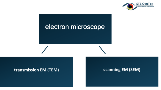
There are two main types of electron microscope – the transmission EM (TEM) and the scanning EM (SEM). Our laboratory is equipped with a state-of-the-art Zeiss TEM900 electron microscope, and this is used together with a number of subsidiary techniques (e.g. immunogold labelling, negative staining) to examine particular issues. Our highly experienced personnel can perform the entire procedure for you, right through from the initial preparation to the final analysis.


