OcuTox has great experience with histology and histopathology at the light and electron microscopical level of eye tissues from humans, monkeys, pigs cattle, dogs, rats and mice. Particularly the electron microscopy is a prodigious tool that can be effectively be used in mode of action and toxicity/safety studies.
We also perform labelling of the proteins by immuno-gold technique at an ultrastructural level

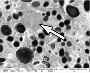
Immuno-gold labelling of a phagosome in an RPE cell from a rat

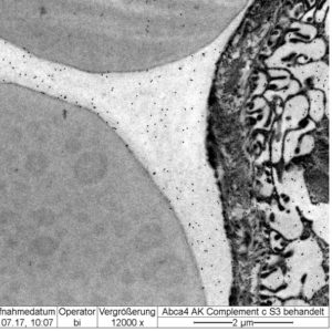
Immuno-gold labelling of complement factors in the serum of a choriocapillaris and basal labyrinth of an RPE cells from a mouse
and follow the distribution of drugs by light microscopical

A light microscopical autoradiogram shows the penetration of a radio labelled drug through the fovea of a monkey. The drug becomes visible by induction of silver grains (arrowheads). Within the fovea the penetration is different (arrow) compared to the parafovea (arrowheads)
and ultrastructural autoradiography

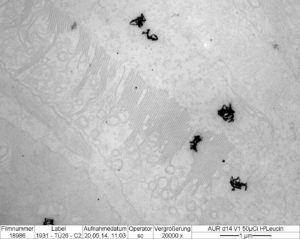
An electron microscopical autoradiogram shows the uptake of a radio labelled drug into a cone photoreceptor outer segment of a monkey

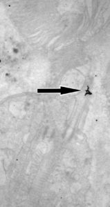
An electron microscopical autoradiogram shows the transport of a radio labelled drug through the cilium of a cone photoreceptor outer segment from a monkey

