Ocular neurodegeneration models
- Retinal ganglion cell (RGC) loss in optic nerve clamping model
- RGC loss in model of elevated intraocular pressure (high IOP or HIOP)
- Photoreceptor apoptosis in animal models of blue light damage (LD) retinal degeneration
- Genetically modified mice and rats
- Light toxicity
Applications
- Dry AMD
- Geographic atrophy
- Glaucoma
- Central retinal artery or vein occlusion (CRAO, CRVO)
Evaluations
- Electroretinography (ERG)
- Histology RPE or retinal outer nuclear layer (ONL) thickness
- Ocular Coherence Tomograohy (OCT).
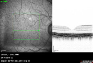
SLO with corresponding OCT image through the fovea of a monkey
Immuno-cytochemistry