Mouse or rat models with retinoblastoma are available

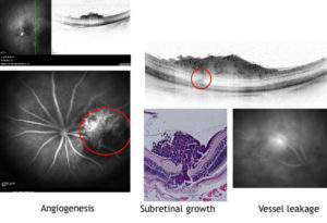
Ocular tumor modell in a nude mouse,the red circle left shows the blood vessel of the tumor in an angiogram . OCT of the same site shows penetration of the tumor through the retina (red circle right) by OCT and corresponding histology of the same site (lower row middle). The tumor vessels leak out fluorescein (lower row right)

